
1Orthogonal Research and Education Lab, 6270 Canterbury Dr Zionsville, IN 46077, USA. Email: shussainather@gmail.com
2Gulf Specimen Marine Laboratory & Aquarium, 222 Clark Drive Panacea, FL 32346, USA.
*Corresponding author : Richard Gordon
Gulf Specimen Marine Laboratory & Aquarium, 222 Clark Drive Panacea, FL 32346 USA.
Email: DickGordonCan@protonmail.com
Received: Sep 28, 2024
Accepted: Oct 22, 2024
Published Online: Oct 29, 2024
Journal: Journal of Artificial Intelligence & Robotics
Copyright: © Gordon R (2024). This Article is distributed under the terms of Creative Commons Attribution 4.0 International License.
Citation: Ather SH, Gordon R. Electronic steering of X-rays. J Artif Intell Robot. 2024; 1(2): 1012.
This study presents a novel approach to electronically steering X-rays using Janus spheres arranged in multiple layers, each reflecting at an example 4o Bragg angle. Utilizing 11 layers of these dual-material spheres, our method aims to achieve rapid x-ray steering, significantly reducing radiation dose, particularly in early breast cancer detection, via new CTR algorithms. Additionally, we review potential applications of this technology in x-ray astronomy and microscopy.
The field of medical imaging, particularly in the detection of early-stage breast cancer, faces significant challenges with current technologies. Conventional mammography often faces challenges such as low resolution, higher than necessary radiation exposure, and reliance on 2D projections, which are particularly problematic for detecting small, early-stage tumors. To address these challenges, our research focuses on the development of Ray-by-Ray Computed Tomography (RBRCT), which minimizes x-ray dose to normal tissue using steerable X-ray beams. Although initial prototypes may employ an industrial robot for steering a polycapillary x-ray source, this paper proposes the use of Janus spheres for enhanced speed. The primary goal of this system is to enhance imaging precision, reduce radiation doses, and ablate premetastasis lesions, allowing >99% predicted cure rate [1-3,5,7-19]. Here we propose the use of Janus spheres, specially designed dual-material spheres capable of reflecting X-rays at precise angles. This configuration enables electronic steering of X-rays, allowing for controlled redirection of the beam to improve imaging accuracy and minimize dose [1]. Our design incorporates 11 layers of Janus spheres, each reflecting x‑rays at a 4o angle, creating a staggered pattern that maximizes coverage and steering range up to +44o from a parallel x-ray beam Note that 4o is not exceptional, as NaCl has a Bragg angle of about 7o [2]. With possibly 95% Bragg reflection efficiency {Bajt, 2012 #887100] [17], 11 layers could still transmit .9511 = .57 or 57%, depending on the material composition and layer structure. This supports our design choice of using Janus spheres configured at a 4o Bragg angle to maintain high reflection efficiency. Thus, we include a movable collimator to protect the patient from the up to 43% of scattered x-rays. The Imatron [12] was the first example of electronic steering, first proposed in 1950 [11], in its case, via magnetic steering of an electron beam impinging on an x-ray target. In our case, we directly steer an x-ray beam. While robotic x-ray steering is demonstrable with a robot carrying a parallel polycapillary x-ray source, electronic steering of the x-rays from such a source would be much faster.
Janus sphere configuration
Janus spheres are arranged in multiple layers to achieve steering of X-rays through electronic rotation of each sphere. Each Janus sphere layer reflects X-rays at a consistent 4-degree angle, and the configuration of 11 layers was chosen keep scattering losses under 50%. These layers are staggered to cover a broad range of angles, enhancing the overall beam steering capability. The Janus spheres are electronically addressable, which allows for dynamic control over the beam. This setup provides a significant improvement in directing X-rays to specific regions of interest, especially useful in complex imaging scenarios like breast cancer detection. All of the Janus spheres in a given row would be rotated to the same angle for a parallel beam of x-rays (They would have to be rotated individually if the beam converged or diverged). We have not addressed the rotation mechanism here. Electronic paper using rotating Janus spheres was originally designed to replace newsprint with a binary display [14,23]. It is presently used for assorted book readers [22].
Simulation of reflection of X-rays off Janus spheres
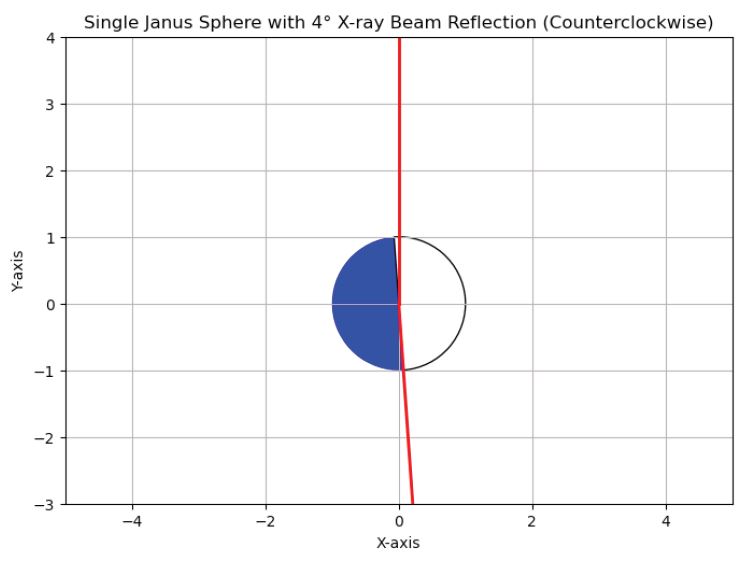
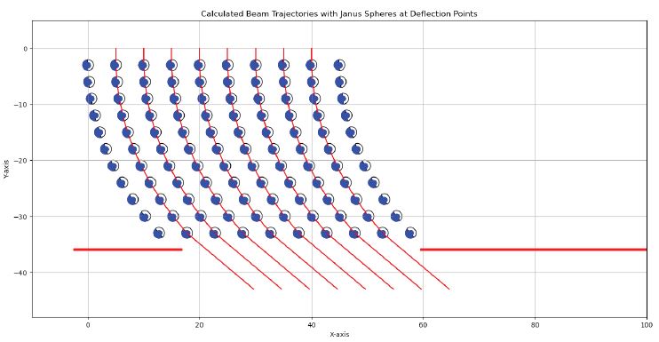
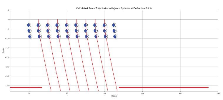
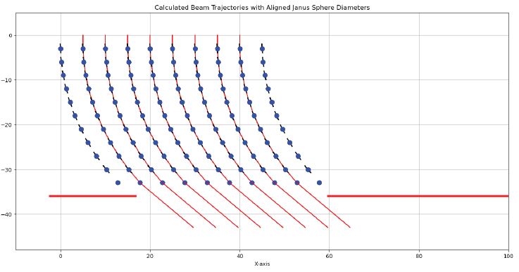
Whereas it is practical, but slow, to aim a parallel polycapillaryx-ray source (2 kg) via an industrial robot, we would like to do so more quickly. The basic idea is to bounce an x-ray microbeam at a low glancing angle corresponding to the Bragg angle of the mirror (Figure 1). We propose configuring the Janus sphere as a mirror, with one hemisphere transparent to x-rays and the other reflective, to achieve controlled beam steering. Each Janus sphere in consecutive layers would be rotated to deflect the beam another increment. With a Bragg angle of 4o and 11 layers, we could deflect the beam up to +44o. The remaining photons would be lost to scattering and absorption. They could for the most part be blocked from entering the patient using a movable sheet of x-ray opaque material with an elliptic hole (collimator) that permits passage of the parallel beam. This concept is illustrated in Figure 2.
If the Janus spheres in a given layer are placed adjacent to one another, their center-to-center displacement is 2r, then in a linear array the spheres would intercept a fractional area of = 78.5%. The lost x-rays would be absorbed or scattered and most blocked by the collimator.
For lower angles than +44o, the lower rows of Janus spheres could be slid out of the way of the beam, as in Figure 3, allowing angles that are multiples of 4o.
The problems to be solved are:
1. Design and manufacture of a material with the maximum possible Bragg angle and minimum loss;
2. Rotating each Janus sphere to the appropriate angle (the same for all spheres in a given layer) within the peak reflection around the Bragg angle, perhaps involving a feedback mechanism;
3. Overcoming rotational Brownian motion of the possibly nanoparticle Janus spheres (again possibly by feedback).
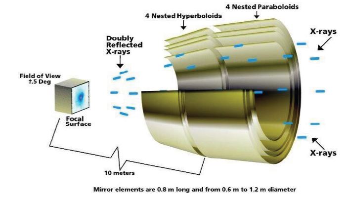
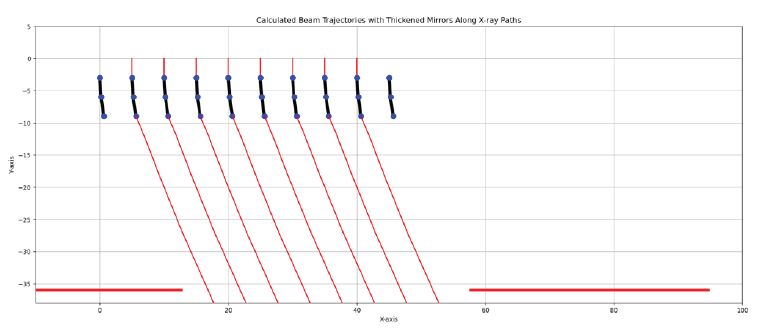
As the X-ray signal is received via detector pixels, those may be used as a source of feedback (albeit of multiple parameters) and filtering out remaining scattered X-rays. Macroscopic feedback systems for Bragg angles have been developed [18]. Note that an alternative is to use multiple two-sided sheets of a reflecting material. This would be analogous the multilayered x-ray telescopes (Figure 4), but not fixed, such as adaptive mirrors for x-ray microscopy [10]. Our cascading design of Janus spheres or mirrors apparently has not been proposed elsewhere [3,5-9,15,16,19,24]. However, explicit piezoelectric steering mechanisms proposed might be miniaturizable.
Here we have adapted mirrors to directing a parallel beam of x-rays in Figure 5 and Figure 6.
With multifocal breast tumors, images acquired at intervals may help in their early detection [13].
Traditional methods, such as fan or cone beam Computed Tomography (CT), involve higher radiation doses due to irradiation of all normal tissues with the same x-ray intensity as directed at potential tumors. Attempts have previously been made to tailor CT to the patient. With steered x‑ray beams, such as proposed here, the dose to normal tissues could be substantially reduced. This requires new Computed Tomography (CT) algorithms, such as RBYRCT (Ray By Ray CT), which are under development, involving a modified MART algorithm, machine learning inpainting, and halting once tumors are detected, minimizing dose. In the context of 3D breast CT, the use of compression paddles could subsequently be used to ablate small, premetastasis tumors without having to relocate them. Overall, our approach is “search & destroy”, predicted to result in >99% cure rate of such tumors [20].
The cumulative error is analogous to the situation for the “cranes on cranes” model for morphogenesis of Daniel Dennett [4,6]. And therefore places a limit on the number of rows of Janus spheres and the steering that could be accomplished. However, feedback mechanisms that compensate for angular and positional errors for individual Janus spheres may be possible, so that manufacturing errors could be tolerated. We leave those problems for future engineers, but see that in principle, electronic steering of X-rays is plausible. It should also be much faster than robotic steering. The concept of steering x-rays is not new; early ideas date back to foundational work in the mid‑20th century [11]. However, the practical implementation of such systems was historically limited by technological constraints, including the lack of suitable materials and control systems. Recent advancements in nanotechnology, coupled with the development of parallel polycapillary X-ray sources, have brought this vision [21] closer to reality. The integration of modern computational techniques and materials science makes it plausible to achieve precise, real-time control of x-ray beams, potentially transforming the landscape of medical imaging.
Optimization of Janus sphere materials: Future research could explore the development of new materials or coatings for Janus spheres or mirrors to enhance x-ray reflectivity and minimize absorption. This may involve experimenting with various materials to achieve the optimal balance between reflection efficiency and structural stability.
Advanced feedback mechanisms: Integrating more sophisticated feedback mechanisms is essential for real-time adjustments to Janus sphere positioning and orientation. Developing systems that can dynamically monitor and adjust the configuration of Janus spheres in response to patient movement or changes in beam intensity would significantly improve image quality and accuracy.
Integration with machine learning: Implementing machine learning algorithms to predict and correct for distortions in real-time could further enhance the precision of RBRCT imaging. This would involve training models on a range of imaging scenarios to develop predictive capabilities for system calibration and optimization.
Expansion to other medical applications: Beyond breast cancer detection, exploring the use of RBRCT in other medical fields such as interventional radiology, neurology, or oncology could open new avenues for research and clinical application.
Prototype development and clinical trials: Moving from theoretical and computational models to physical prototypes is crucial. Conducting initial clinical trials would provide valuable data on the system’s performance in real-world settings and help refine both the technology and its applications.